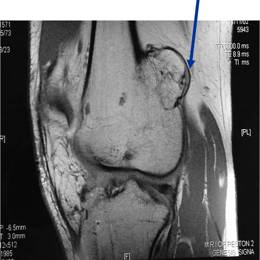
Osteochondroma

The most common benign tumor, which constitutes 35% of all benign bone tumors, and 10% of all bone tumors overall. It is an overgrowth of the bone that happens at the end of the bone near the growth plate. It is made up of both bone and cartilage. 

Osteochondroma9
Screen Shot 2022 08 19 At 11.26.50 AM

What is an Osteochondroma?

Osteochondromas are outgrowths that occur at the end of the bone near the growth plate and are covered with cartilage. The prevalence is unknown because many are asymptomatic and found accidentally on radiographs (X-rays, MRIs, etc.) taken for other reasons. The most common benign tumor, which constitutes 35% of all benign bone tumors, and 10% of all bone tumors overall.

Who is usually affected?
  • • Male predominance 3:1. 
  • • Usually presents in the third decade of life.
  • • Most commonly between ages 10 and 30.
Common Bones Involved
  • • Any portion of the skeleton that has cartilage can develop the condition.
  • • Most common in the knee area (35% of cases).
Causes
Causes
  • • There is a autosomal dominant inheritance genetic cause.
Signs and Symptoms
Signs and Symptoms
  • • Signs and symptoms include pain and a hard swelling.
Biological Behavior
Biological Behavior
  • • Osteochondromas are benign, or noncancerous tumors, so they will not spread to other parts of the body. 


Diagnosis
Diagnosis
  • • The work-up often consists of a physical examination, X-rays, CT scans, MRI, and sometimes bone scans are required. CT scans can be used to check for subtle mineralization that may help with the diagnosis
  • • CT of the chest is necessary to check for pulmonary metastases. The lungs and other bones are the to most common sites for the tumor to spread.
  • • The diagnosis is often confirmed with a biopsy, which means taking a sample of tumor and having it analyzed under a microscope by a pathologist.

Risk to your limbs

Osteochondromas are noncancerous but aggressive tumors that, if left unchecked, will grow and destroy your normal bone and invade the surrounding tissues.

Radiographic imaging is used to help form a diagnosis. These include X-Ray, MRI, CT and Bone Scans

An example of an Osteochondroma MRI is shown.

Osteochondroma15b

Treatment of

Osteochondroma

Treatment includes simple excision of the tumor. In most cases, no treatment is required other than regular monitoring. Recurrence after surgery is rare.

Intralesional Curettage

Intralesional Curettage means to scoop the tumor out using a spoon-like tool called a curette. This is a surgery that aims to remove the mass and restore the bone so that the patient can get back to normal function. The ABC is identified within the bone and scooped, or curetted, out. The cavity is then shaved down with a Midas Rex Drill, which is similar to a dental drill. This drill removes more tumor cells.

Bone Grafting and Fixation

The empty bone cavity is usually filled with bone graft or bone cement. Bone can be donated (allograft) or taken from the patient themselves (autograft). Fixation devices, such as a plate and screws, may be used in specific situations to prevent postoperative fracture. 

I've seen many doctors and I can confidently attest Dr. Wittig is the preeminent orthopaedic specialist. He is genuinely kind and caring, as he demonstrated by completely addressing my concerns and compassionately relating to what I was dealing with. He clearly outlined the plan of attack, and recommended the two additional doctors who would become part of my 'team'. Dr. Wittig was so effective in allaying our fears and bringing us optimism. My surgery was significant, but I was up and walking the next day and back at the gym 5 weeks later. This is further testament to Dr. Wittig's skill. He saved my leg and my life, and I feel so very blessed to say he is my doctor. I have already recommended him to others, and I will continue to do so. I would trust him with my closest family and lifelong friends. BEST DOCTOR EVER.

S.G.

Img16516

Hey! I am Dr. Wittig.

Myself and my amazing team are dedicated to saving your life and your limb. Losing a limb because of a tumor can be a terrifying experience. But, it does not have to be the only option. I’ve spent 20+ years as a Board-Certified Orthopedic Surgeon and Orthopedic Oncologist.

I’ve devoted my career to helping children and adults afflicted with bone and soft tissue masses by performing complex limb saving surgeries. Most patients can have their limb saved, which may require innovative techniques.

Patients afflicted with musculoskeletal tumors have complex conditions that are best taken care of at large hospitals. I am the Chairman of Orthopedics and Chief of Orthopedic Oncology at Morristown Medical Center. My philosophy is a multidisciplinary team approach, working together to tailor treatment to individual patients. Education and research are essential to my practice, providing the best setting for extraordinary patient care. Because of this, we have some of the top results in the country.