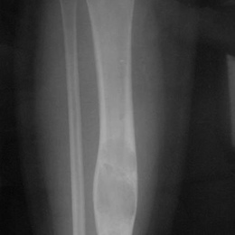
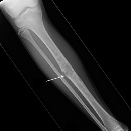
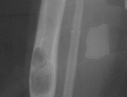
Adamantinoma

Adamantinomas are rare, slow-growing cancerous bone tumors that form primarily in the tibia, also known as the shin bone, but occasionally form in the jaw, forearm, hands or feet. In 20% of cases, the tumor travels to the lungs or the lymph nodes.

Adamantinoma 01
Screen Shot 2022 10 05 At 4.26.50 PM

What is an Adamantinoma?

An adamantinoma is a rare, slow-growing bone sarcoma (cancer). It might arise from a pre-existing condition from the bone called an osteofibrous dysplasia. Since these tumors are slow-growing, they rarely spread to other sites in the body.

Who is usually affected?
  • • Most common in 20 to 40 year olds.
  • • Peak age is in the third decade of life. 
  • • Males and females are affected equally.


Causes
Causes
  • • There is a recurrent pattern of numerical abnormalities including extra copies of chromosomes 7, 8, 12, 19 and 21. 
  • • There is a chromosomal translocation. 
  • • Many are thought to to arise from osteofibrous dysplasia. 
Common Bones Involved
  • • Most commonly occurs in the center of the shinbone, which is known as the tibia bone. 
  • • Occasionally in the jaw, forearm, hands or feet.
  • • This includes the fibula, ulna, femur, humerus and radius. 


Signs and Symptoms
Signs and Symptoms
  • • Symptoms include a slow-growing swelling with or without pain.
  • • A break due to the tumor weakening the bone.
  • • When the tumor develops specifically in the tibia, the lower leg may have a deformed appearance due to curving (bowing) of the bone. 
  • • Bone deformity and fracture due to the cancer are the main reasons patients seek medical attention. 
  • • 50% of cases have a history of localized trauma.
Biological Behavior
Biological Behavior
  • • Over 80% of these tumors are at least 5 cm in size.
  • • There are two types: Classic vs. Differentiated. 
  • • Classic type grows beyond the bone cortex, is seen in older patients, and sometimes metastasizes, or spreads to other areas of the body. 
  • • Differentiated type is confined to the bone cortex, seen in earlier age, and does not metastasize.
  • • Sarcomas are categorized into low-grade, intermediate-grade or high-grade lesions based on a variety of biological factors. This grade classification represents their biologic aggressiveness and correlates with the likelihood of the cancer spreading to other parts of the body.


Diagnosis
Diagnosis
  • Biopsy: The diagnosis of musculoskeletal tumors is based on clinical evaluation, radiological imaging, and pathological findings from a biopsy. A biopsy is when the surgeon cuts out a piece of the tumor and looks at it under a microscope. This is important for confirmation of the diagnosis for accurate classification before creating a definitive treatment plan. Open incisional biopsy is the most reliable of current available diagnostic methods. 
  • Imaging: Staging studies for a sarcoma include plain radiographs (X-rays), computed tomography (CT), and magnetic resonance (MRI) scans. X-Ray remains the key imaging in evaluating a bone lesion. The CT scan allows the doctor to understand the extent of the tumor. The MRI scan provides information on the tumors relation to vital structures, including nerves and blood vessels. Further imaging may be needed including CT of the chest and positron emission tomography (PET) scans to rule out the spread of the cancer to other parts of the body. Sometimes a test called a bone scan is ordered, which is a whole-body method of understanding the entire skeleton. 

Risk to your limbs

Adamantinomas are cancerous tumors that, if left unchecked, will grow and destroy your normal bone. As the tumor grows, the bone is weakened and you are at an increased risk of breaking the bone due to the tumor, called a pathological fracture.

Radiographic imaging is used to help form a diagnosis of ABC. These include X-Ray, MRI, CT and Bone Scans

An example of an Adamantinoma X-Ray is shown.

98A) Plain AP X Ray Of Right Tibia Adamantinoma

I've seen many doctors and I can confidently attest Dr. Wittig is the preeminent orthopaedic specialist. He is genuinely kind and caring, as he demonstrated by completely addressing my concerns and compassionately relating to what I was dealing with. He clearly outlined the plan of attack, and recommended the two additional doctors who would become part of my 'team'. Dr. Wittig was so effective in allaying our fears and bringing us optimism. My surgery was significant, but I was up and walking the next day and back at the gym 5 weeks later. This is further testament to Dr. Wittig's skill. He saved my leg and my life, and I feel so very blessed to say he is my doctor. I have already recommended him to others, and I will continue to do so. I would trust him with my closest family and lifelong friends. BEST DOCTOR EVER.

S.G.

Img16516

Hey! I am Dr. Wittig.

Myself and my amazing team are dedicated to saving your life and your limb. Losing a limb because of a tumor can be a terrifying experience. But, it does not have to be the only option. I’ve spent 20+ years as a Board-Certified Orthopedic Surgeon and Orthopedic Oncologist.

I’ve devoted my career to helping children and adults afflicted with bone and soft tissue masses by performing complex limb saving surgeries. Most patients can have their limb saved, which may require innovative techniques.

Patients afflicted with musculoskeletal tumors have complex conditions that are best taken care of at large hospitals. I am the Chairman of Orthopedics and Chief of Orthopedic Oncology at Morristown Medical Center. My philosophy is a multidisciplinary team approach, working together to tailor treatment to individual patients. Education and research are essential to my practice, providing the best setting for extraordinary patient care. Because of this, we have some of the top results in the country.