A total scapula prosthetic replacement is a procedure that involves removing a tumor, which usually malignant or benign aggressive, of the scapula/shoulder girdle and replacing the shoulder with a special customizable total shoulder prosthesis called total scapula prosthesis with a proximal humerus component. It is often used to reconstruct the shoulder after limb sparing surgery for a sarcoma arising from or involving the scapula.
The shoulder girdle, as seen in the figure below, consists of the proximal humerus, scapula, and clavicle. The shoulder joint is formed by the glenoid (cup), which is part of the scapula, and the proximal humerus ball, which is the upper portion of your arm. There are many muscles that attach to your scapula, including the rotator cuff muscles. The scapula, also known as your shoulder blade, is a common site for primary sarcomas to arise. Some of these include chondrosarcoma, osteosarcoma and Ewing’s sarcoma. Soft tissue sarcomas can also develop around the scapula and shoulder, and invade the adjacent bones. Limb-sparing surgery can be performed for approximately 95% of tumors arising from or around the shoulder girdle. In order to reconstruct the shoulder girdle with a total scapula prosthesis one must be able to preserve the deltoid muscle and axillary nerve, trapezius muscle, rhomboids and latissimus. In some instances, the extremity cannot be saved, and a forequarter amputation is performed.
Contraindications for saving the limb may include neurovascular invasion, infection, pathological fracture, invasion of the chest wall, extensive disease, contamination from a poorly performed biopsy, recurrent disease.
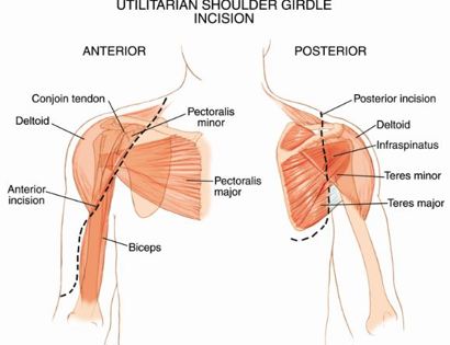
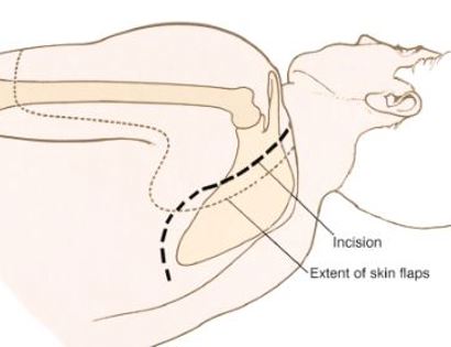
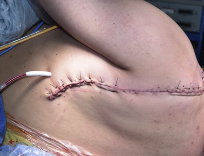
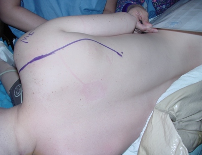
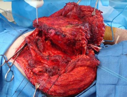
The utilitarian shoulder girdle incision is used. A long, longitudinal incision along the lateral border of the scapula is utilized that may extend over the shoulder and down the arm depending on the size of the mass. The skin is raised up to expose the muscles around the scapula.
Occasionally, a scapular resection can be performed completely through the posterior incision (an incision made through the back); however, if there is a large anterior (frontal) portion of the tumor with displacement of the axillary vessels (vessels located in the underarm), it may be beneficial to also incorporate an anterior incision in the front of the shoulder, which may make it easier to separate the blood vessels and nerves from the tumor.
The rhomboids, trapezius, latissimus, serratus anterior, and deltoid are released from the scapula followed by separating the important blood vessels and nerves. The remaining muscle attachments are then released. Developing surgical planes (margins that are tumor free) and separating muscles that can be preserved and leaving those in continuity with the tumor that should be removed. This is based on preoperative MRI and intraoperative findings as well as the type of tumor. The rotator cuff is also removed with the tumor, as it harbors microscopic cells.
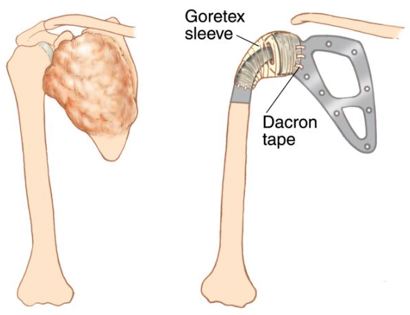
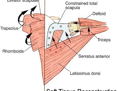
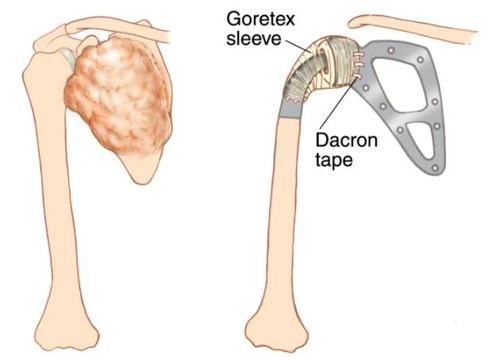
There are two types of methods to reconstruct the limb once the tumor is removed. Reconstruction of the scapula/shoulder joint utilizing a specialized tumor prosthesis is the most common limb-sparing technique. If the surrounding muscles can be saved after the tumor of the scapula has been removed, then a scapular prosthesis can be utilized to restore the defect (space where the scapula was removed). The muscles involved in the reconstruction process include deltoid, trapezius, rhomboids, and latissimus dorsi. If there is not a sufficient amount of muscle remaining in the surrounding area after the tumor has been removed, then the humerus (arm bone) is supported from the clavicle with Dacron tape.
This is an image of a scapular prosthesis. This type of prosthesis can be used if there is enough muscles remaining after the tumor is removed.
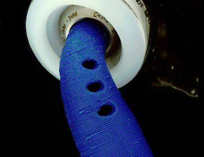
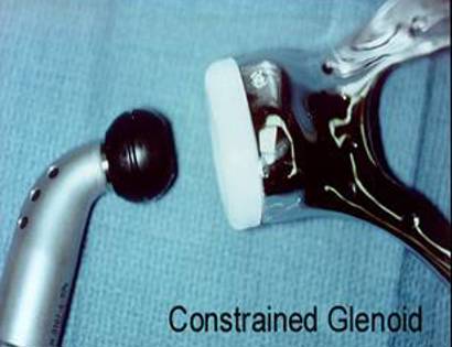
This is a close up of the scapula prothesis joint.
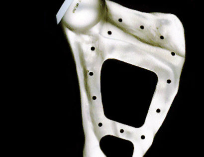
This is a second-generation scapular prosthesis. This allows the muscles a better area to attach in order to give the shoulder more stability.
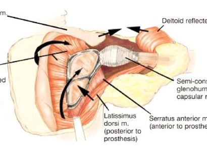
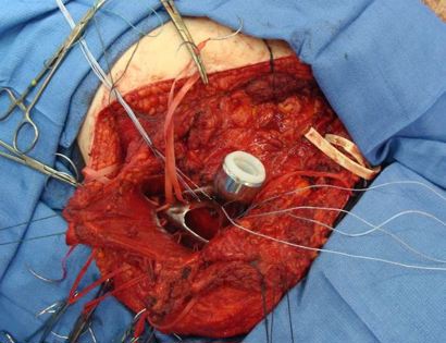
The scapula prosthesis is placed with the upper part of the humerus prosthesis already seated in place. The humerus portion of the prothesis is cemented and then the scapula prosthesis is sutured to the chest wall. This allows for proper positioning and function of the arm.
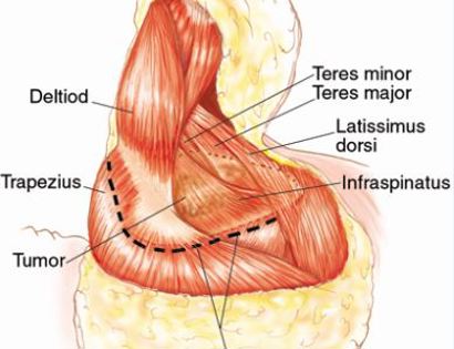
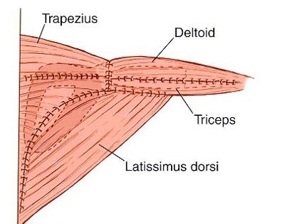
This figure shows the soft-tissues involved in the reconstruction and coverage of the prosthesis.
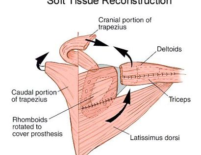
This figure shows the soft-tissue reconstruction.
In cases where muscle reconstruction is a viable option, the proximal humerus (upper arm) and scapula are replaced with prostheses to restore the shoulder joint. The proximal humerus prosthesis is cemented into the bone and the scapula prosthesis is secured to the chest wall by sutures. The latissimus dorsi, deltoid, and trapezius can then be utilized to cover over the prostheses and help fill in the defect.
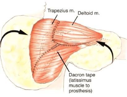
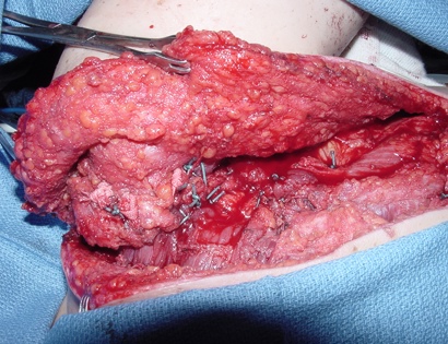
A series of muscles including the trapezius, deltoid, triceps, and latissimus dorsi are used to cover the entire scapula prosthesis.
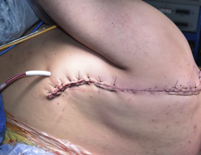
The incision is closed with sutures and bandages are placed to cover the surgical site. Multiple large drains may also be inserted and are used to drain the surgical site and prevent seroma (buildup of fluid).
This is an example of a scapula/shoulder blade tumor as seen on an MRI.
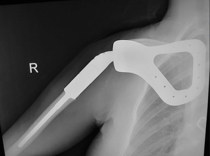
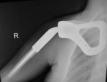
This image shows the total scapula prosthetic replacement as seen on an X-ray.
This is an image of the incision utilized in a total scapula prosthetic replacement.
This is an image of the scapula being removed from the chest wall.
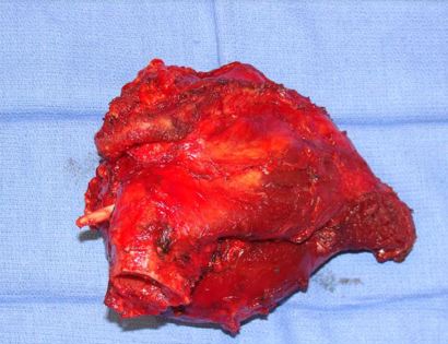
This is an image of the specimen with the tumor removed in its entirety.
This image shows the scapula prosthesis inserted after the tumor, scapula, and margins have been removed.
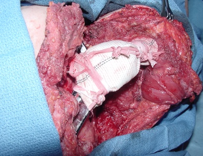
In this image, ligaments are reconstructed with a dacron tube (surgical tape) and heavy sutures.
This is an image of the soft tissue fully encompassing the scapula prosthesis.
This image shows the complete closure of the total scapula prosthetic replacement, with the drain intact.
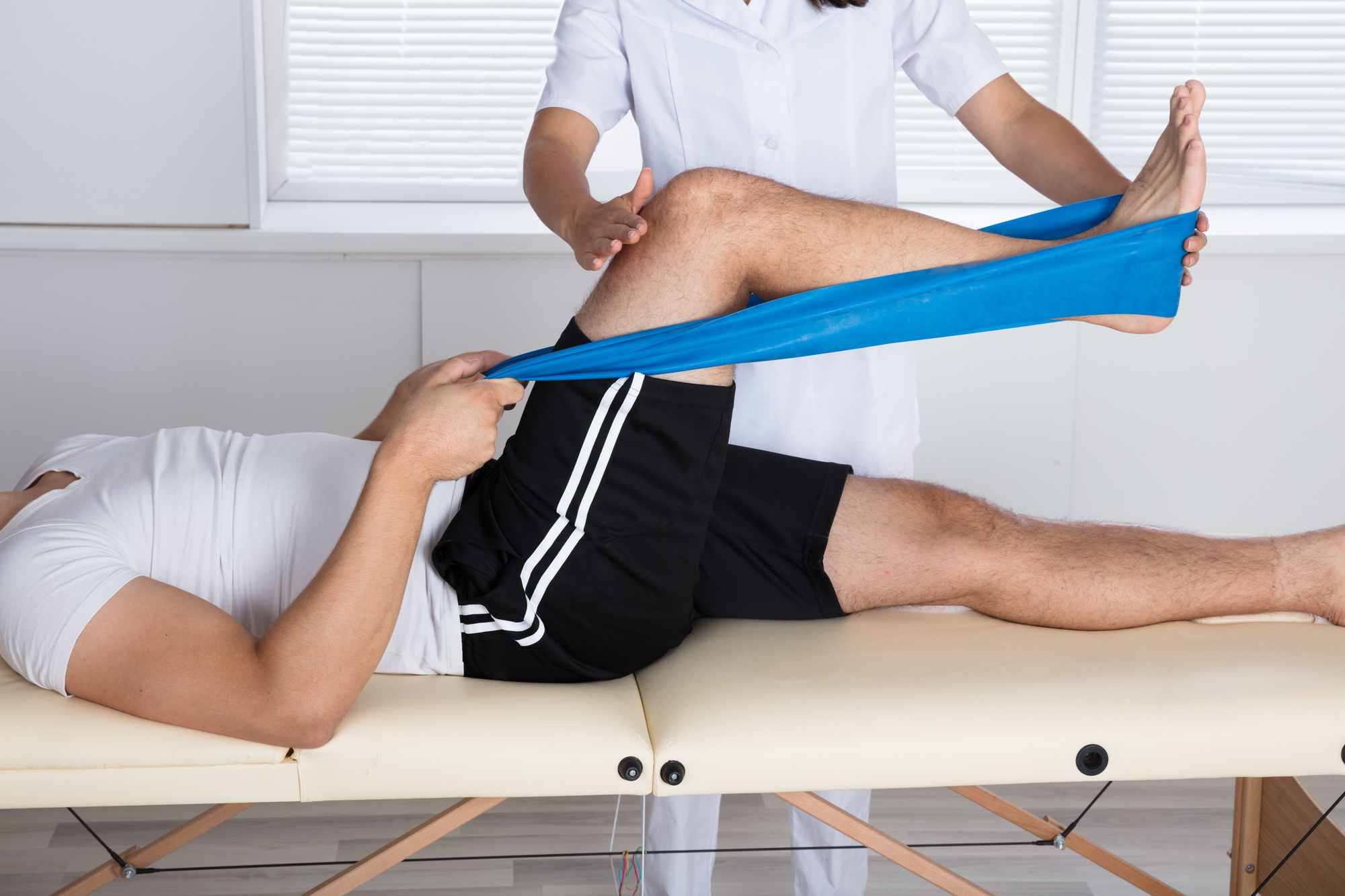
After your surgery you will spend a few nights in the hospital and then will be recuperating at home or at a rehabilitation center. Various pain protocols and nerve blocks are used to minimize pain. Mostly, all patients are very comfortable after the surgery. For the first few days you will ice the area and keep it elevated to reduce swelling. You will return to the office 2 weeks after surgery for a post-operative visit. Patients are usually kept in a sling for 6 weeks to allow the muscles to heal. The elbow is not permitted to extend beyond 45 degrees of flexion for 4 to 6 weeks to allow the biceps muscle to heal which will stabilize the prosthesis. Once cleared, you will subsequently start physical therapy. We usually prescribe specific physical therapy protocols 3 times a week for 12 weeks after surgery to gradually strengthen muscles. Strengthening with significant resistance after sufficient range of motion is achieved as determined by Dr. Wittig. There may be an ultimate weight limit imposed upon you depending on various factors.
You will be monitored periodically with X-rays over the course of 5 years. Sometimes an MRI and/or CT may be used to additionally monitor the area to make sure the tumor has not come back. You will then have follow up appointments every 4 months for the first 2 years, then every 6 months for the next 2 years, and then once a year. Since the bone integrity has been restored to full or almost full, recovery is anticipated provided the patient adheres to strict physical therapy.
Dr. James Wittig narrates a video illustrating the surgical technique for a total scapula prosthetic replacement surgery. | WATCH VIDEO